ACL Tear vs MCL Tear Differences & Treatment
Acl tear vs mcl tear what is the difference and treatment options – ACL tear vs MCL tear: what is the difference and treatment options? That’s the burning question for anyone facing knee pain after an injury. These two ligaments, the anterior cruciate ligament (ACL) and the medial collateral ligament (MCL), are crucial for knee stability, and tearing one can be seriously debilitating. Understanding the differences between an ACL and MCL tear—from their symptoms and diagnosis to the various treatment options available—is key to a successful recovery.
This post dives deep into both injuries, helping you navigate the complexities and empowering you to make informed decisions about your health.
We’ll explore the anatomy of each ligament, compare common causes, delve into the diagnostic process (including physical exams and imaging), and Artikel the treatment pathways—both surgical and non-surgical—for each. We’ll also cover rehabilitation and recovery, providing insights into what to expect during your journey back to full functionality. Think of this as your comprehensive guide to understanding and managing ACL and MCL tears.
Introduction to ACL and MCL Tears
Understanding the differences between an ACL tear and an MCL tear is crucial for proper diagnosis and treatment. Both injuries affect the knee joint, but they involve different ligaments and result in distinct symptoms and recovery pathways. This section will delve into the anatomy and function of these ligaments, clarifying their roles in knee stability and outlining common causes of injury.
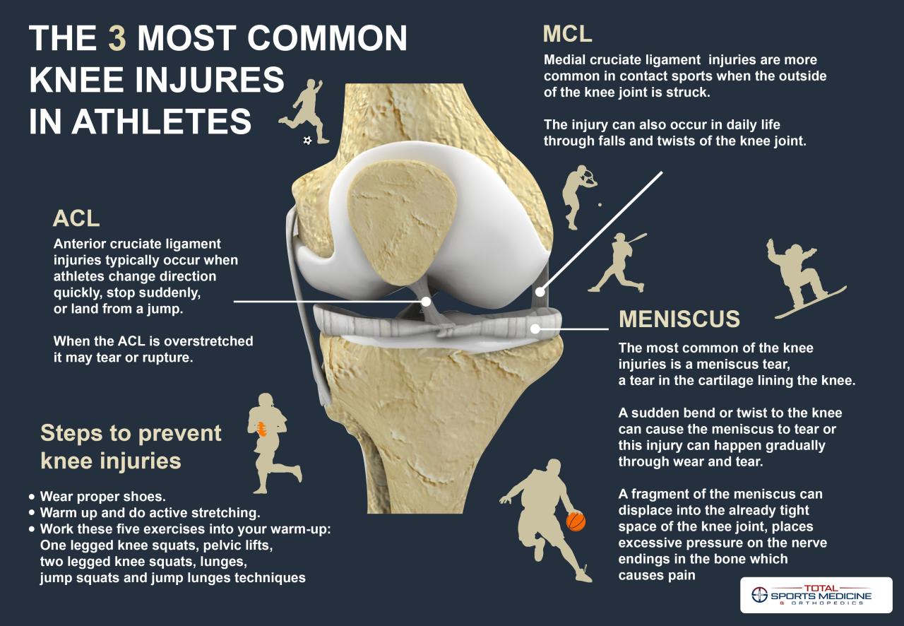
An ACL tear is a rupture or stretching of the anterior cruciate ligament (ACL), a crucial stabilizer of the knee joint. This ligament prevents the tibia (shinbone) from sliding out in front of the femur (thighbone), particularly during twisting or sudden stopping motions.
An MCL tear, on the other hand, involves damage to the medial collateral ligament (MCL), located on the inner side of the knee. The MCL’s primary function is to provide stability against outward forces, preventing the knee from bending excessively inward.
ACL and MCL Anatomy and Function
The ACL and MCL, while both vital for knee stability, have distinct anatomical locations and functional roles. The ACL is positioned deep within the knee joint, connecting the femur to the tibia. Its primary function is to restrict anterior tibial translation – preventing the shinbone from moving too far forward relative to the thighbone. Conversely, the MCL is located on the outer surface of the knee joint, running along the inner side of the knee.
Its role is to resist valgus stress, which is a force pushing the knee inward. Imagine trying to force your knees together – that’s the kind of stress the MCL protects against. While both ligaments work in concert to maintain knee stability, they are susceptible to injury through different mechanisms.
Causes of ACL and MCL Tears
The causes of ACL and MCL tears often overlap, but some mechanisms are more strongly associated with one injury than the other. Direct blows to the knee can cause either injury, but the type of force and the resulting stress on the ligaments will determine which one tears.
| Cause | ACL Tear | MCL Tear | Notes |
|---|---|---|---|
| Direct Blow | Often involves a blow to the outside of the knee, causing hyperextension and internal rotation | Often involves a blow to the outside of the knee, causing valgus stress | Both can result from direct impact. |
| Non-Contact Injury | Common in sports involving sudden changes in direction, jumping, and landing, often involving twisting or deceleration. | Less common than in ACL tears, but can occur with similar mechanisms involving forceful inward stress on the knee. | These injuries are often caused by biomechanical factors. |
| Hyperextension | Significant hyperextension can damage the ACL. | Can also damage the MCL but less frequently than with valgus stress. | Overextension of the knee joint. |
| Valgus Stress | Less directly involved but can contribute to overall instability leading to injury. | Directly involved, often causing a stretch or tear of the ligament. | Force pushing the knee inwards. |
Symptoms of ACL and MCL Tears

Source: pinimg.com
Understanding the symptoms of an ACL (Anterior Cruciate Ligament) and MCL (Medial Collateral Ligament) tear is crucial for accurate diagnosis and appropriate treatment. While both injuries affect the knee, they present with distinct characteristics, allowing for differentiation. The severity of symptoms can vary depending on the extent of the tear.
ACL Tear Symptoms
An ACL tear often results in a sudden, popping sensation in the knee, followed by immediate pain and swelling. The instability of the knee joint is a hallmark symptom, making it difficult to bear weight or continue activity. Other common symptoms include: a feeling of the knee “giving way,” knee stiffness, and limited range of motion.
Bruising around the knee may also develop over time. The severity of these symptoms can range from mild discomfort to debilitating pain, depending on the nature and extent of the tear. For example, a partial tear might cause less immediate swelling and instability than a complete rupture.
MCL Tear Symptoms
In contrast to an ACL tear, an MCL tear often presents with pain along the inner side of the knee. Swelling is usually present, but may not be as immediate or severe as with an ACL tear. Instability is often less pronounced than in an ACL tear, although significant MCL tears can cause noticeable knee instability. The pain is typically exacerbated by activities that stress the medial side of the knee, such as twisting or direct blows to the inner knee.
ACL and MCL tears are common knee injuries, with ACL tears often requiring surgery while MCL tears sometimes heal conservatively. The advancements in medical technology are incredible; for instance, I just read about the FDA approving clinical trials for pig kidney transplants in humans – fda approves clinical trials for pig kidney transplants in humans – which is amazing progress! Getting back to knee injuries, choosing the right treatment for your specific tear depends on factors like the severity and your individual needs.
The ability to bear weight may be only mildly affected, especially in less severe tears. For instance, a grade I MCL sprain (minor stretching) might only cause mild pain and minimal swelling, whereas a grade III MCL tear (complete rupture) could cause significant instability and pain.
Comparing and Contrasting Symptoms
The following bullet points highlight the key differences and similarities in the symptoms of ACL and MCL tears:
- Popping Sensation: More common in ACL tears.
- Immediate Swelling: More pronounced and rapid in ACL tears.
- Instability: More significant and debilitating in ACL tears; often less severe in MCL tears, although severe MCL tears can still cause instability.
- Pain Location: ACL tears often cause pain in the center of the knee or slightly anterior, while MCL tears cause pain along the inner (medial) aspect of the knee.
- Mechanism of Injury: ACL tears frequently occur during sudden twisting or hyperextension movements, while MCL tears often result from direct blows to the outer side of the knee, causing valgus stress (force pushing the knee inward).
- Weight-bearing Ability: Often significantly impaired immediately after an ACL tear, less so in MCL tears (unless severe).
Diagnostic Flowchart
A flowchart visualizing the diagnostic process based on symptoms would begin with the initial presentation of knee pain and swelling following a traumatic event. The first branching point would ask: “Is there a significant popping sensation at the time of injury?” A “yes” would lead down a path focusing on ACL tear characteristics (significant instability, rapid swelling, pain in the center/anterior knee), while a “no” would lead to a branch focusing on MCL tear characteristics (pain on the inner knee, less severe instability, potentially less rapid swelling).
Further branching could incorporate assessment of specific movements (e.g., valgus stress test for MCL) and range of motion to further refine the diagnosis. Ultimately, the flowchart would converge on a provisional diagnosis, requiring confirmation through imaging (MRI) and physical examination. This visual aid would simplify the decision-making process for clinicians, guiding them through a systematic approach to differentiating between ACL and MCL injuries based on the patient’s reported symptoms.
Diagnosis of ACL and MCL Tears: Acl Tear Vs Mcl Tear What Is The Difference And Treatment Options
Diagnosing ACL and MCL tears requires a combination of a thorough physical examination and, in most cases, imaging studies. The accuracy of diagnosis significantly impacts the treatment plan and subsequent recovery. While some injuries are immediately obvious, others require a more nuanced approach to differentiate between the two and rule out other potential causes of knee pain.
Physical Examination for ACL Tears
The physical exam for an ACL tear focuses on assessing the stability of the knee joint. The most common tests include the Lachman test, anterior drawer test, and pivot shift test. The Lachman test involves gently pulling the tibia forward while the knee is slightly flexed. A significant amount of anterior tibial translation indicates a potential ACL tear.
The anterior drawer test is similar, but the knee is flexed to approximately 90 degrees. Again, excessive forward movement of the tibia suggests an ACL injury. The pivot shift test assesses the rotational stability of the knee and is more complex, often performed by experienced clinicians. Positive findings in these tests strongly suggest an ACL tear, but further investigation is usually needed for confirmation.
Physical Examination for MCL Tears
Diagnosing an MCL tear primarily involves assessing the medial aspect of the knee. The valgus stress test is the cornerstone of this examination. With the knee extended and then flexed to 30 degrees, the examiner applies a valgus force (pushing the knee inward). Pain and increased laxity (excessive movement) in the medial joint line point towards an MCL injury.
The severity of the injury is often graded based on the degree of laxity felt during this test. Tenderness to palpation along the medial collateral ligament also supports the diagnosis.
Role of Imaging in ACL and MCL Tear Diagnosis
Imaging plays a crucial role in confirming the diagnosis and assessing the extent of the injury. X-rays are typically performed initially to rule out fractures or other bony abnormalities. However, X-rays are not ideal for visualizing soft tissues like ligaments. Magnetic resonance imaging (MRI) is the gold standard for visualizing ligamentous injuries. MRI provides detailed images of the ACL and MCL, allowing clinicians to assess the integrity of the ligaments and identify any associated injuries such as meniscus tears or cartilage damage.
MRI is particularly useful in differentiating between partial and complete tears.
Diagnostic Accuracy of Various Methods
| Diagnostic Method | ACL Tear Accuracy | MCL Tear Accuracy | Notes |
|---|---|---|---|
| Physical Examination (Lachman, Anterior Drawer) | Sensitivity: 70-90%, Specificity: 80-95% | Sensitivity: Varies, Specificity: Varies | Accuracy depends on examiner experience; less reliable for MCL than ACL. |
| Valgus Stress Test | Low | Sensitivity: 60-80%, Specificity: 70-90% | Primarily used for MCL; less useful for ACL. |
| MRI | Sensitivity: >90%, Specificity: >90% | Sensitivity: >90%, Specificity: >90% | Gold standard; provides detailed visualization of ligamentous structures. |
| X-ray | Low (rules out fractures) | Low (rules out fractures) | Not useful for visualizing ligaments; primarily for ruling out bone injuries. |
Treatment Options for ACL Tears

Source: ytimg.com
An ACL tear, depending on the severity, requires careful consideration of treatment options. The decision between surgical and non-surgical approaches hinges on factors like the patient’s age, activity level, and the extent of the tear. Both options aim to restore knee stability and function, but the path to recovery differs significantly.
Non-Surgical Treatment for ACL Tears
Non-surgical treatment is often considered for patients with partial ACL tears, minor injuries, or those with low activity levels who are not heavily reliant on their knee for athletic pursuits. This approach focuses on managing pain and swelling, improving knee stability, and restoring function through conservative methods. It usually involves a combination of strategies to help the knee heal naturally.
Surgical Treatment for ACL Tears
Surgical reconstruction is typically recommended for complete ACL tears, especially in younger, more active individuals who participate in sports or activities demanding significant knee stability. The goal of surgery is to replace the torn ligament with a graft, restoring the knee’s structural integrity.
ACL Reconstruction Surgical Techniques
Several surgical techniques are available for ACL reconstruction. The choice depends on factors like the surgeon’s preference, the patient’s anatomy, and the extent of the injury. Common techniques include using a graft from the patient’s own body (autograft), such as a hamstring tendon or patellar tendon, or a graft from a donor (allograft). The surgeon will drill tunnels in the tibia and femur, then secure the graft in place using specialized instruments.
The procedure is typically performed arthroscopically, meaning minimally invasive incisions are made, leading to smaller scars and quicker recovery times.
Rehabilitation After ACL Surgery, Acl tear vs mcl tear what is the difference and treatment options
Rehabilitation is a crucial part of recovering from ACL surgery and regaining full knee function. The rehabilitation process is usually divided into phases, each focusing on specific goals. The initial phase, often lasting several weeks, focuses on pain management, reducing swelling, and regaining range of motion. This involves physical therapy sessions focusing on gentle exercises and range-of-motion activities.
The next phase focuses on strengthening the muscles around the knee and improving stability. Progressive weight-bearing and more advanced exercises are introduced gradually. The final phase involves a return to sports and activities, with a focus on regaining pre-injury athletic performance levels. The entire process can take several months, sometimes even a year or more, depending on individual factors and the complexity of the surgery.
Comparison of Treatment Options for ACL Tears
| Treatment Option | Pros | Cons | Suitable For |
|---|---|---|---|
| Non-Surgical Treatment | Avoids surgery, less invasive, quicker initial recovery | May not be effective for complete tears, longer recovery time to full function, potential for instability | Partial tears, low activity levels, older individuals |
| Surgical Reconstruction (Autograft) | High success rate for restoring stability, good long-term results, suitable for high-impact activities | Invasive procedure, longer recovery time, potential for complications like infection or graft failure, requires extensive rehabilitation | Complete tears, young, active individuals, athletes |
| Surgical Reconstruction (Allograft) | Faster rehabilitation in some cases, avoids harvesting a graft from the patient | Potential for rejection, higher cost, slightly higher risk of complications | Complete tears where autograft is not feasible |
Treatment Options for MCL Tears
Medial collateral ligament (MCL) tears, unlike ACL tears, often heal without surgery. The treatment approach depends heavily on the severity of the tear and the individual’s activity level. Conservative management is usually the first line of defense, with surgery reserved for only the most severe cases.
Non-Surgical Treatment for MCL Tears
Non-surgical treatment focuses on reducing pain, inflammation, and restoring function. This typically involves a combination of rest, ice, compression, and elevation (RICE), along with physical therapy. The initial phase emphasizes reducing swelling and protecting the joint. As healing progresses, physical therapy focuses on regaining range of motion, strength, and stability. This might include exercises to strengthen the muscles surrounding the knee, improve proprioception (awareness of the joint’s position in space), and gradually return to normal activities.
The duration of non-surgical treatment varies depending on the severity of the tear and the individual’s response to therapy, but it can range from several weeks to several months. Pain management may involve over-the-counter medications like ibuprofen or naproxen, or prescription pain relievers in some cases.
Surgical Treatment for MCL Tears
Surgical intervention for MCL tears is relatively uncommon. Surgery is typically considered only when there’s a complete tear of the MCL, combined with damage to other knee ligaments (like an ACL or PCL tear), or when non-surgical treatments have failed to provide adequate healing and restoration of function after a reasonable timeframe. The surgical procedure usually involves repairing the torn ligament, often using sutures to reattach the torn ends.
In some cases, reconstruction might be necessary, but this is less frequent than with ACL tears. The choice between repair and reconstruction depends on the specific nature of the injury and the surgeon’s assessment. For example, a complete tear with significant retraction might require reconstruction, while a partial tear with minimal displacement might be amenable to repair.
Rehabilitation Following MCL Injury
Rehabilitation following an MCL tear, whether treated surgically or conservatively, is crucial for a full recovery. The rehabilitation program is tailored to the individual’s needs and the severity of the injury. Early stages focus on pain and swelling management, restoring range of motion, and protecting the knee joint. As healing progresses, the focus shifts to strengthening the muscles around the knee, improving balance and proprioception, and gradually increasing activity levels.
ACL and MCL tears are common knee injuries, differing in location and resulting instability. Understanding the distinctions is crucial for effective treatment, which can range from physical therapy to surgery. It’s amazing how focused we get on physical health sometimes; I recently learned about managing completely different challenges, like those faced by families dealing with Tourette Syndrome in children, which requires a very different approach as detailed in this helpful article: strategies to manage tourette syndrome in children.
Returning to knee injuries, proper diagnosis is key for choosing the right path towards recovery from an ACL or MCL tear.
A physical therapist guides the patient through a carefully designed program of exercises, progressing in intensity and complexity as the knee heals. This might involve exercises like range-of-motion exercises, strengthening exercises (using weights or resistance bands), balance exercises, and functional exercises that mimic activities of daily living. Return to sports or high-impact activities is typically a gradual process, guided by the patient’s progress and the physical therapist’s assessment.
Comparison of ACL and MCL Rehabilitation Protocols
The rehabilitation process for ACL and MCL injuries differs significantly in duration, intensity, and focus.
- Duration: ACL rehabilitation typically takes longer (6-12 months or more) than MCL rehabilitation (several weeks to months), due to the complexity of the injury and the need for complete ligament healing.
- Focus: ACL rehabilitation emphasizes regaining knee stability and preventing future injuries, often requiring intensive strengthening and proprioceptive training. MCL rehabilitation primarily focuses on restoring range of motion, reducing pain and swelling, and regaining muscle strength.
- Intensity: ACL rehabilitation often involves a more rigorous and structured program, with a gradual progression to high-impact activities. MCL rehabilitation is generally less intense, with a quicker return to low-impact activities.
- Surgical Intervention: Surgical intervention is much more common for ACL tears than for MCL tears.
Recovery and Rehabilitation
Recovering from an ACL or MCL tear requires a dedicated rehabilitation program. The time it takes to return to full activity varies greatly depending on several factors, and successful recovery hinges on diligent adherence to the prescribed plan. This section will detail those factors, and provide examples of exercises commonly used in physical therapy for both injuries.Recovery time for both ACL and MCL tears is influenced by numerous factors.
These include the severity of the tear (partial vs. complete), the individual’s age and overall health, the presence of other injuries, the adherence to the rehabilitation protocol, and the individual’s pre-injury fitness level. For example, a young, healthy athlete with a partial MCL tear and excellent compliance with physical therapy might recover within a few weeks, while an older individual with a complete ACL tear and other underlying health conditions could require several months, or even longer.
Furthermore, the speed of recovery is also dependent on the individual’s commitment to the rehabilitation process and their ability to consistently perform exercises correctly.
Factors Influencing Recovery Time
Several key factors determine the length of the recovery process. Age plays a significant role, with younger individuals generally recovering faster due to their body’s natural healing capabilities. The severity of the tear itself is another crucial factor; a complete tear requires more extensive repair and rehabilitation than a partial tear. The presence of concomitant injuries, such as meniscus tears or cartilage damage, can significantly prolong recovery.
Finally, the patient’s compliance with the prescribed rehabilitation program and their overall level of fitness prior to the injury are critical determinants of the recovery timeline. A patient’s commitment to the rehabilitation program, including regular attendance at therapy sessions and diligent home exercise, is crucial for successful recovery.
Physical Therapy Exercises for ACL Tear Rehabilitation
The initial phase of ACL rehabilitation focuses on reducing pain and swelling, regaining range of motion, and strengthening the muscles surrounding the knee. As the knee heals, the focus shifts towards regaining stability and improving neuromuscular control.
- Straight Leg Raises: Lying on your back, slowly lift one leg straight up, keeping your knee straight and your leg aligned with your hip. This strengthens the quadriceps.
- Hamstring Curls: Lying on your stomach, slowly bend your knee, lifting your heel towards your buttock. This strengthens the hamstrings.
- Isometric Quadriceps Sets: While lying on your back with your leg straight, tighten your thigh muscle by pushing your kneecap against the surface beneath it. Hold for several seconds, then relax.
- Mini-Squats: Stand with your feet shoulder-width apart, and perform partial squats, only lowering yourself a few inches. This builds strength and improves stability.
- Balance Exercises: Stand on one leg, focusing on maintaining balance. Progress to more challenging exercises, such as standing on a balance board or foam pad.
Physical Therapy Exercises for MCL Tear Rehabilitation
MCL tear rehabilitation focuses on reducing pain and inflammation, restoring range of motion, and strengthening the supporting muscles of the knee. Emphasis is placed on controlled movements to prevent further injury and promote healing.
So, you’re wondering about ACL vs. MCL tears? The key difference lies in which ligament is affected – the anterior cruciate or medial collateral. Treatment often involves surgery for ACL tears, while MCL tears sometimes heal conservatively. It’s crucial to remember that even seemingly minor injuries can have serious long-term consequences; understanding the risk factors that make stroke more dangerous highlights the importance of proactive health management, just as early diagnosis and appropriate treatment are vital for both ACL and MCL injuries to prevent further complications.
Getting the right diagnosis and treatment plan for your knee injury is key to a full recovery.
- Range of Motion Exercises: Gently bend and straighten your knee, gradually increasing the range of motion as tolerated. This improves flexibility and reduces stiffness.
- Isometric Exercises: These exercises involve contracting the muscles around the knee without moving the joint. This strengthens the muscles without putting stress on the injured ligament.
- Straight Leg Raises (Modified): Similar to ACL rehab, but with a focus on controlled movements and avoiding excessive stress on the MCL.
- Short Arc Quadriceps Exercises: This exercise focuses on strengthening the quadriceps muscle while minimizing stress on the MCL. The knee is bent to a shallow angle (around 30 degrees) during the exercise.
- Light Resistance Exercises: Once the pain subsides, light resistance exercises can be introduced using resistance bands or light weights to further strengthen the leg muscles.
Potential Complications
Complications associated with ACL and MCL injuries and their treatments can include persistent pain, instability, osteoarthritis, stiffness, and infection (in the case of surgery). In some cases, the initial treatment may not be successful, necessitating further intervention. For example, an ACL reconstruction might fail, requiring a revision surgery. Similarly, an MCL tear that doesn’t heal properly can lead to chronic instability and pain, requiring additional treatment such as bracing or further surgery.
Arthritis is a long-term potential complication of both injuries, particularly if there is significant damage to the articular cartilage.
Illustrative Examples
Understanding the differences between ACL and MCL tears is best illustrated through real-world examples. These examples will highlight the typical mechanisms of injury, presenting symptoms, diagnostic approaches, and common treatment pathways for each ligament.
ACL Tear Case Study
Consider a female basketball player, 22 years old, who experiences a sudden twisting injury to her right knee while landing awkwardly from a jump shot. She reports an immediate popping sensation followed by intense pain and rapid swelling. She is unable to bear weight on the leg. Diagnosis, via MRI, confirms a complete tear of the anterior cruciate ligament (ACL).
The treatment plan involves surgical reconstruction using a hamstring autograft, followed by a rigorous physiotherapy program focused on regaining range of motion, strength, and stability. The surgery aims to replace the torn ligament with a graft, restoring knee joint stability. Post-surgery, the athlete will undertake a comprehensive rehabilitation plan, likely involving bracing, physical therapy, and gradual return-to-sport protocols.
MCL Tear Case Study
A 30-year-old male soccer player sustains a direct blow to the outer side of his left knee during a tackle. He feels a sharp pain on the inner side of his knee and experiences immediate instability, though he can still bear some weight. There is less swelling compared to the ACL tear case. Physical examination reveals tenderness along the medial collateral ligament (MCL), and an MRI confirms a grade II MCL sprain (partial tear).
Treatment involves non-surgical management including bracing to provide support and stability, physical therapy focusing on range of motion exercises and strengthening, and gradual return to activity. Surgery is rarely required for MCL tears, unless the injury is severe or accompanied by other ligament damage.
Knee Joint Visual Representation
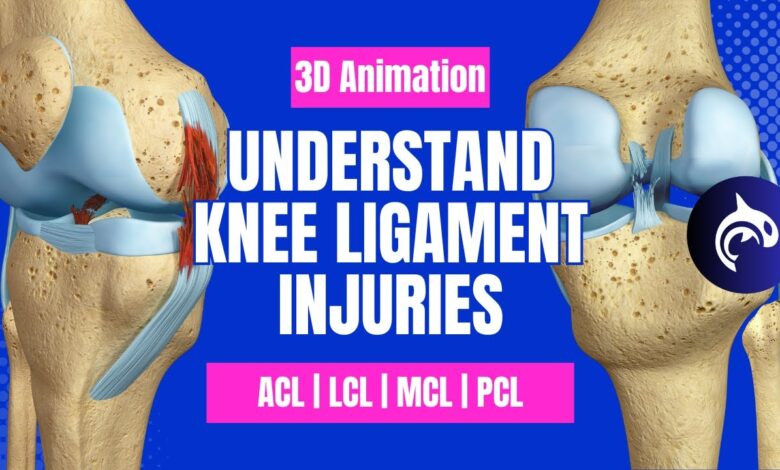
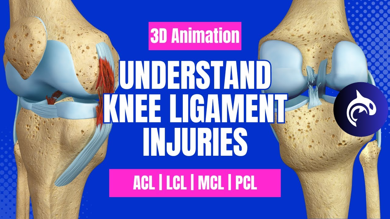
Imagine the knee joint as a hinge, connecting the thigh bone (femur) to the shin bone (tibia). The patella (kneecap) sits in front. The ACL is a strong, cord-like ligament located inside the knee joint, running diagonally from the back of the femur to the front of the tibia. It acts as a primary stabilizer, preventing the tibia from sliding forward relative to the femur.
The MCL is a broader, flatter ligament located on the inner side of the knee, running from the inner side of the femur to the inner side of the tibia. It provides stability to the inner knee, preventing excessive inward movement. Both ligaments work together with other structures (menisci, other ligaments, and joint capsule) to ensure the knee functions smoothly and remains stable.
Last Recap
Navigating an ACL or MCL tear can feel overwhelming, but armed with knowledge, you can approach your recovery with confidence. Remember, understanding the unique characteristics of each injury—from symptoms to treatment—is crucial for a successful outcome. While this post provides a solid overview, always consult with your doctor or physical therapist for personalized advice and a tailored treatment plan.
Your journey back to a healthy, active life starts with informed decisions and consistent effort. Let’s get you moving again!
Questions and Answers
What are the long-term effects of an untreated ACL tear?
Untreated ACL tears can lead to chronic instability, increased risk of further injury (like meniscus tears), and premature arthritis.
Can I return to sports after an MCL tear?
Yes, most people can return to sports after an MCL tear, but the timeline depends on the severity of the injury and the adherence to physical therapy.
How long does it take to recover from ACL surgery?
Recovery from ACL surgery is a lengthy process, typically taking 6-9 months or longer, depending on individual factors and the rehabilitation program.
Is MCL surgery always necessary?
No, MCL tears often heal conservatively with physical therapy. Surgery is usually only considered for severe or unstable tears.
