Doctors Perform Groundbreaking Cataract Surgery on 31-Week-Old Preemie
Doctors perform groundbreaking cataract surgery on thirty one week old premature baby – Doctors perform groundbreaking cataract surgery on thirty-one-week-old premature baby – it’s a headline that grabs you, right? This incredible medical feat pushes the boundaries of pediatric ophthalmology, highlighting the incredible advancements in surgical techniques and neonatal care. Imagine the challenges: operating on an eye so tiny and delicate, a body so fragile, a life so new. This story delves into the specifics of the procedure, the pre- and post-operative care, and the technological marvels that made this success possible.
Prepare to be amazed by the resilience of this little one and the skill of the medical team.
We’ll explore the unique challenges of performing cataract surgery on a premature infant, comparing it to adult procedures and detailing the specialized instruments and techniques involved. We’ll also delve into the ethical considerations, the meticulous pre-operative assessments, and the intensive post-operative monitoring required to ensure the baby’s well-being. The story is a testament to human ingenuity and the unwavering dedication to saving and improving young lives.
The Medical Procedure
Performing cataract surgery on a 31-week-old premature infant presents unique challenges compared to adult procedures. The delicate nature of the infant’s eye structures, coupled with their immature physiological systems, necessitates specialized techniques and instrumentation. The goal is to remove the cataract, preserving as much of the developing eye’s structure and function as possible to maximize the chances of good vision development.The surgery itself involved a meticulous approach.
The surgeon likely used a microscope with a high magnification capability to visualize the tiny structures within the baby’s eye. Specialized micro-instruments, far smaller and more delicate than those used in adult cataract surgery, were employed to make incisions and manipulate the lens. The phacoemulsification technique, commonly used in adult cataract surgery, might have been modified or avoided altogether, given the risk of damaging the delicate tissues in a premature infant’s eye.
Instead, a technique involving aspiration or manual removal of the lens might have been preferred. The choice of technique is heavily influenced by the specific characteristics of the cataract and the overall health of the baby’s eye. Post-operatively, the baby would have received close monitoring and likely required medication to prevent infection and inflammation.
Challenges Posed by the Infant’s Prematurity
The primary challenges in performing cataract surgery on a premature infant stem from their underdeveloped physiological systems and the fragility of their ocular structures. The infant’s small size necessitates the use of specialized, miniature instruments. Maintaining a stable surgical field is significantly more difficult due to the infant’s inability to cooperate and the need for general anesthesia. The immature immune system also increases the risk of infection and complications.
Furthermore, the developing eye’s anatomy is less robust than that of an adult, increasing the potential for inadvertent damage during the procedure. The longer-term implications of the surgery on the developing eye need careful consideration, including the potential need for future interventions. Accurate assessment of the surgical outcome requires specialized equipment and ongoing monitoring to track the baby’s visual development.
Comparison of Cataract Surgery Procedures, Doctors perform groundbreaking cataract surgery on thirty one week old premature baby
The following table highlights key differences between cataract surgery in adults and premature infants:
| Procedure Step | Adult Surgery | Premature Infant Surgery | Rationale for Difference |
|---|---|---|---|
| Anesthesia | Local or regional anesthesia often sufficient | General anesthesia typically required | Infant’s inability to cooperate and maintain stillness. |
| Instrumentation | Standard-sized instruments | Specialized micro-instruments | Infant’s smaller eye size and delicate structures. |
| Surgical Technique | Phacoemulsification commonly used | Aspiration or manual lens removal may be preferred | Reduced risk of damage to delicate tissues in premature infants. |
| Post-operative Care | Relatively straightforward | Intensive monitoring and management of potential complications | Increased risk of infection and other complications in premature infants. |
| Lens Implantation | Intraocular lens (IOL) implantation is common | IOL implantation may be delayed or not performed immediately | The ongoing development of the eye in premature infants necessitates a cautious approach to IOL placement. Potential for future adjustments may be considered. |
Pre-Operative Care and Assessment

Source: toiimg.com
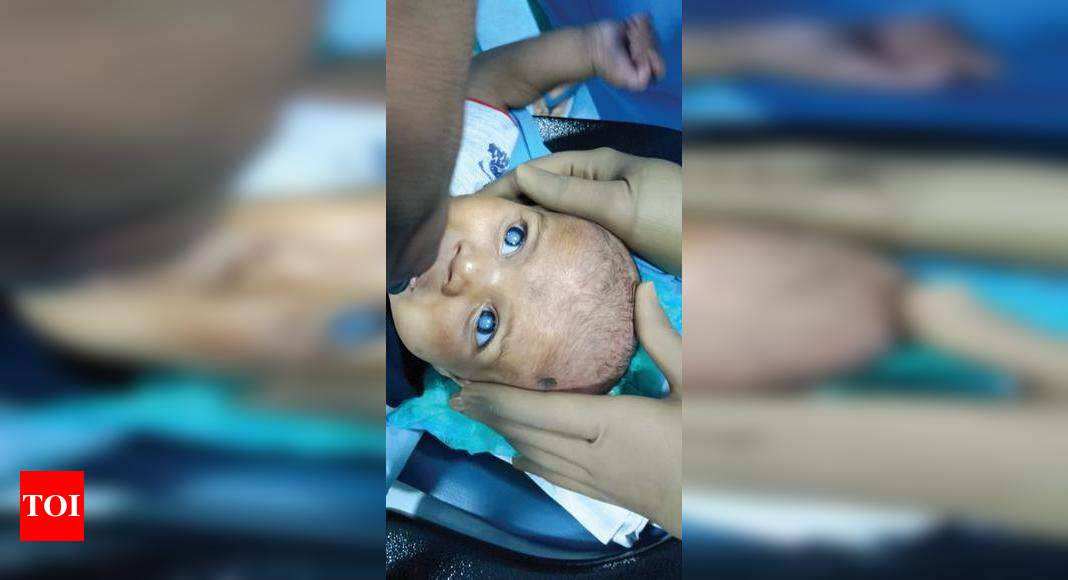
Preparing a 31-week premature infant for cataract surgery presents unique challenges. The fragility of the infant, coupled with their underdeveloped organ systems, necessitates a meticulous and comprehensive pre-operative assessment and preparation strategy. This process aims to minimize risks and optimize the chances of a successful surgical outcome while prioritizing the baby’s well-being.The pre-operative phase involves a multidisciplinary approach, bringing together neonatologists, ophthalmologists, anesthesiologists, and nurses.
Each specialist plays a crucial role in ensuring the infant is as stable as possible before undergoing the procedure. This collaborative effort is critical given the high-risk nature of operating on such a young patient.
Pre-Operative Assessments and Preparations
Thorough pre-operative assessment is paramount for ensuring the safety and success of the surgery. This includes a detailed review of the infant’s medical history, focusing on their gestational age, birth weight, current health status, and any existing medical conditions. Furthermore, the infant’s vital signs, including heart rate, respiratory rate, blood pressure, and oxygen saturation, are closely monitored.
The ophthalmological assessment focuses on the severity and location of the cataracts, the presence of any associated eye conditions, and the overall visual acuity.A critical aspect is optimizing the infant’s physiological stability before surgery. This might involve adjusting respiratory support, managing any underlying infections, and ensuring adequate nutrition and hydration. For example, if the infant is on respiratory support, the anesthesiologist might need to adjust ventilator settings to optimize gas exchange during the procedure.
Similarly, if the infant is experiencing feeding difficulties, adjustments to nutritional support might be necessary to maintain energy levels and overall stability.
- Complete medical history review, including gestational age and birth weight.
- Comprehensive ophthalmological examination to assess cataract severity and location.
- Assessment of respiratory function and oxygen saturation levels.
- Evaluation of cardiovascular status, including heart rate and blood pressure.
- Assessment of nutritional status and hydration levels.
- Blood tests to check for infections and other potential complications.
- Electrocardiogram (ECG) to assess heart function.
- Imaging studies (if necessary) to rule out other abnormalities.
Anesthesiology in Premature Infants
Anesthesiology plays a pivotal role in managing the risks associated with surgery in premature infants. The primary concern is the immature organ systems, which are more susceptible to the effects of anesthesia. The anesthesiologist must carefully select appropriate anesthetic agents and techniques, considering the infant’s gestational age, weight, and overall health status. The goal is to provide adequate analgesia and anesthesia while minimizing potential complications such as respiratory depression, bradycardia, and hypotension.
Close monitoring of vital signs and the infant’s response to anesthesia is crucial throughout the procedure. Specialized monitoring equipment, such as pulse oximetry and capnography, are essential for detecting any adverse events promptly. The anesthesiologist will frequently adjust medication levels to maintain stable vital signs. For example, they may use lower doses of medication than in adults and titrate them carefully based on the infant’s response.
Ethical Considerations
Performing surgery on a premature infant raises significant ethical considerations. The primary ethical principle is beneficence – the action must be in the best interest of the child. This necessitates a careful weighing of the potential benefits of the surgery (improved vision) against the potential risks and side effects. Parental consent is essential, and it’s crucial that parents are fully informed about the procedure, its risks, and potential benefits.
The decision-making process must involve open communication and shared decision-making between the medical team and the parents. Furthermore, the surgical team must adhere to the highest standards of care, minimizing any potential risks to the infant’s well-being. The potential for long-term complications must be considered and discussed with the parents. For example, potential risks of retinal detachment or other eye complications associated with the surgery are carefully weighed against the potential benefits of improved vision.
Post-Operative Care and Monitoring

Source: ytimg.com
The successful completion of cataract surgery on a 31-week-old premature infant is only the first step in a long and delicate journey. Post-operative care is crucial for ensuring the baby’s comfort, preventing complications, and maximizing the chances of a positive visual outcome. This phase demands meticulous attention to detail and a highly specialized approach.Post-operative care for this tiny patient involved a multifaceted approach focusing on pain management, infection prevention, and close monitoring of vital signs and ocular health.
The infant remained in the Neonatal Intensive Care Unit (NICU) under continuous observation for several days following the procedure.
It’s amazing what medical advancements can achieve; doctors successfully performed groundbreaking cataract surgery on a thirty-one-week-old premature baby! This incredible feat highlights the strides in neonatal care, but it also makes me think about the fragility of young lungs. Reading about Monali Thakur’s hospitalization after struggling to breathe, and learning how to prevent respiratory diseases from this article monali thakur hospitalised after struggling to breathe how to prevent respiratory diseases really underscores the importance of preventative care, especially for vulnerable infants.
The successful surgery on the premature baby gives me hope for the future of medicine, and the need for increased awareness regarding respiratory health in newborns is just as crucial.
Pain Management and Medication
Pain management in a premature infant requires a delicate balance. We used a combination of non-pharmacological methods, such as gentle swaddling and kangaroo care (skin-to-skin contact with a parent), alongside pharmacological interventions. The infant received very low doses of acetaminophen, carefully adjusted according to weight and response, to control any post-operative discomfort. Regular assessments of pain levels were conducted using validated neonatal pain scales.
Topical antibiotic eye drops were administered prophylactically to prevent infection.
Post-Operative Monitoring and Potential Complications
Continuous monitoring of the infant’s vital signs (heart rate, respiratory rate, oxygen saturation, temperature) was essential. We closely monitored the surgical site for any signs of infection, bleeding, or inflammation. Regular ophthalmological examinations were performed to assess the healing process and the clarity of the intraocular lens. We also monitored for any signs of retinopathy of prematurity (ROP), a condition that can affect premature infants.
Potential Complications and Risk Mitigation
The following table Artikels potential complications, their likelihood, and mitigation strategies employed:
| Complication | Description | Likelihood (Relative) | Mitigation Strategy |
|---|---|---|---|
| Infection | Bacterial or fungal infection of the eye. | Low (with prophylactic antibiotics) | Prophylactic antibiotic eye drops, meticulous sterile technique during surgery and post-operative care. |
| Bleeding | Hemorrhage within the eye. | Low | Careful surgical technique, close monitoring of vital signs. |
| Increased Intraocular Pressure | Elevated pressure within the eye. | Low | Regular monitoring of intraocular pressure, prompt treatment if elevated. |
| Retinal Detachment | Separation of the retina from the underlying tissue. | Very Low | Careful surgical technique, close monitoring for signs of retinal detachment. |
| Posterior Capsule Opacification | Clouding of the posterior lens capsule. | Moderate (long-term) | YAG laser capsulotomy if necessary in the future. |
Long-Term Monitoring and Follow-Up Care
Long-term follow-up is critical to ensure the continued health and visual development of the infant. Regular ophthalmological examinations are scheduled at intervals determined by the infant’s progress and any identified risk factors. These examinations assess visual acuity, refractive error, and overall ocular health. Early detection of any issues allows for timely intervention and maximizes the chances of achieving the best possible visual outcome.
The frequency of these checkups will gradually decrease as the child grows, but ongoing monitoring will continue throughout childhood.
Technological Advancements
The successful cataract surgery on the 31-week-old premature infant was a testament to significant advancements in ophthalmic technology. Miniaturization, improved imaging, and refined surgical techniques converged to enable a procedure previously considered too risky for such a young and delicate patient. This wasn’t simply a smaller version of adult cataract surgery; it required a completely different approach, made possible only by recent technological breakthroughs.The surgery’s success hinged on several key technological advancements.
These innovations addressed the challenges posed by the baby’s size, underdeveloped eye structures, and the inherent risks associated with operating on such a fragile patient.
It’s amazing what medical advancements are happening! Doctors successfully performed groundbreaking cataract surgery on a thirty-one-week-old premature baby, a testament to the incredible strides in pediatric ophthalmology. This reminds me of another huge leap forward: the FDA recently approved clinical trials for pig kidney transplants in humans, as reported here fda approves clinical trials for pig kidney transplants in humans.
These breakthroughs, both tiny and massive in scale, highlight the incredible potential of medical innovation to improve lives.
Microscopic Surgical Instruments and Techniques
The use of incredibly fine, specialized instruments was paramount. These instruments, significantly smaller than those used in adult cataract surgery, allowed for precise manipulation within the baby’s tiny eye. For instance, phacoemulsification, the technique used to break up and remove the clouded lens, was performed with a micro-phacoemulsification probe capable of generating ultrasonic energy with extreme precision and minimal collateral damage.
The incision itself was likely incredibly small, minimizing trauma and scarring, thanks to advancements in blade technology and surgical techniques focusing on minimal-incision cataract surgery (MICS). These advancements ensured the surgeon could accurately target the cataract while minimizing potential harm to the surrounding delicate tissues.
Advanced Imaging Technologies
Pre-operative assessment was crucial and relied heavily on advanced imaging technologies. High-resolution ultrasound imaging provided detailed images of the baby’s eye structures, allowing the surgical team to accurately assess the cataract’s size, location, and the overall health of the eye. This non-invasive technique is especially valuable in premature infants where the use of bright light, as in traditional ophthalmoscopy, might be damaging.
Intraoperative imaging techniques, such as surgical microscopes with integrated cameras and high-magnification capabilities, also played a vital role in guiding the surgeon during the procedure. Real-time visualization ensured precision and minimized the risk of complications.
Specialized Anesthesia and Monitoring
The success of the surgery also depended on advancements in anesthesia and patient monitoring. The infant required specialized anesthesia tailored to their age and delicate condition, minimizing the risk of respiratory or cardiovascular complications. Continuous monitoring of vital signs, including heart rate, blood pressure, oxygen saturation, and body temperature, was crucial to ensure the baby’s safety throughout the procedure.
Real-time monitoring systems provided immediate feedback to the anesthesia team, allowing for prompt adjustments and proactive management of any potential problems.
Hypothetical Future Advancements: Robotic-Assisted Cataract Surgery
Improvements in robotic surgery could further enhance the safety and effectiveness of pediatric cataract surgery. Imagine a scenario where a minimally invasive robotic system, guided by advanced imaging and AI, performs the surgery with even greater precision and dexterity than a human surgeon. This system could potentially reduce the risk of human error, minimize trauma to the surrounding tissues, and allow for more complex procedures to be performed on even younger or more fragile patients.
Existing robotic surgery systems are already used in adult cataract surgery, but further miniaturization and refinement of these systems, coupled with advancements in AI-driven image analysis, could revolutionize pediatric ophthalmic surgery. For example, a system could automatically compensate for the infant’s breathing movements, enhancing surgical precision and reducing the need for lengthy periods of intubation.
Impact and Significance
The successful cataract surgery on this 31-week premature infant represents a significant leap forward in pediatric ophthalmology. It demonstrates the feasibility of complex eye surgery in extremely premature babies, pushing the boundaries of what was previously considered possible and offering a beacon of hope for countless others. This achievement not only improves the quality of life for this particular infant but also paves the way for refining techniques and expanding access to similar procedures globally.This groundbreaking surgery highlights the ongoing evolution of surgical techniques and the development of specialized instruments designed for the delicate anatomy of premature infants.
The ability to perform such a procedure successfully at this gestational age challenges the established norms and significantly expands the therapeutic options available to neonatologists and ophthalmologists caring for premature babies. The long-term implications for visual development and overall health in these vulnerable infants are profound.
Groundbreaking Procedures in Premature Infants
Past advancements in neonatal care have laid the groundwork for this success. For example, the development of surfactant replacement therapy revolutionized the treatment of respiratory distress syndrome in premature infants, significantly improving survival rates. Similarly, advancements in neonatal intensive care, including improved ventilation strategies and nutritional support, have created a more stable environment for performing complex surgical interventions.
It’s amazing what medical advancements can achieve! Doctors successfully performed groundbreaking cataract surgery on a thirty-one-week-old premature baby, a testament to the incredible strides in neonatal care. Thinking about the future of these little ones, it made me recall a recent article about Karishma Mehta getting her eggs frozen and the risks involved , highlighting the importance of reproductive choices and planning for the future.
The contrast between these two stories underscores the remarkable possibilities and challenges in modern medicine.
The increasing sophistication of imaging techniques, such as high-resolution ultrasound, also allows for more precise pre-operative planning and assessment.
Potential Implications for Future Treatment
This successful surgery holds immense potential for the future treatment of premature infants with similar conditions. It opens the door to earlier intervention for congenital cataracts, potentially minimizing the impact on visual development and reducing the long-term risks associated with delayed treatment. Furthermore, the refined surgical techniques and specialized instruments employed in this case can be adapted and improved for use in other pediatric ophthalmological procedures.
This includes surgeries for retinopathy of prematurity (ROP), another significant cause of vision impairment in premature infants. The improved understanding of the delicate balance between surgical intervention and the infant’s fragile system gleaned from this experience is invaluable.
Illustrative Description of the Surgery
Imagine a surgical field no larger than a dime. The infant’s eye, tiny and exquisitely fragile, rests within a specialized surgical cradle designed to maintain its delicate position and minimize pressure. The surgeon, wielding instruments finer than a sewing needle, carefully manipulates the lens, its clouded opacity a stark contrast to the surrounding healthy tissues. Microscopic forceps, designed with almost imperceptible gripping surfaces, are used to delicately remove the cataractous lens.
The precision required is extraordinary; a single slip could result in irreparable damage. The entire procedure is performed under a high-powered operating microscope, magnifying the surgical field many times over, allowing the surgeon to navigate the intricate anatomy with utmost care. Every movement is deliberate, every incision calculated, reflecting the surgeon’s years of training and experience in managing such a high-stakes procedure.
The miniature size of the instruments, coupled with the magnification, underscores the extraordinary technical challenge presented by this surgery. The surgeon’s steady hand, unwavering focus, and meticulous technique are essential to the successful outcome.
Final Conclusion: Doctors Perform Groundbreaking Cataract Surgery On Thirty One Week Old Premature Baby
The successful cataract surgery on this thirty-one-week-old premature baby stands as a beacon of hope, showcasing the remarkable progress in pediatric ophthalmology. It’s a story not just of medical triumph, but of resilience, innovation, and the unwavering commitment to providing the best possible care for our most vulnerable patients. This case underscores the importance of continued research and development in neonatal care and highlights the potential for even more groundbreaking advancements in the future.
It leaves us pondering the incredible possibilities that lie ahead in the world of pediatric medicine and the transformative power of medical innovation.
Detailed FAQs
What are the long-term visual outcomes for babies who undergo this type of surgery?
Long-term visual outcomes vary depending on several factors, including the severity of the cataract, the timing of surgery, and the overall health of the baby. Regular follow-up appointments are crucial to monitor vision development and address any potential complications.
What are the risks associated with anesthesia in such a young patient?
Anesthesia in premature infants carries inherent risks, including respiratory complications and cardiovascular instability. However, advancements in anesthetic techniques and careful monitoring significantly reduce these risks.
How common is cataracts in premature babies?
Cataracts are relatively uncommon in premature infants, but they can occur and often require early intervention to prevent vision impairment.
