What is a Full Body Scan for Cancer Detection and Is It Safe?
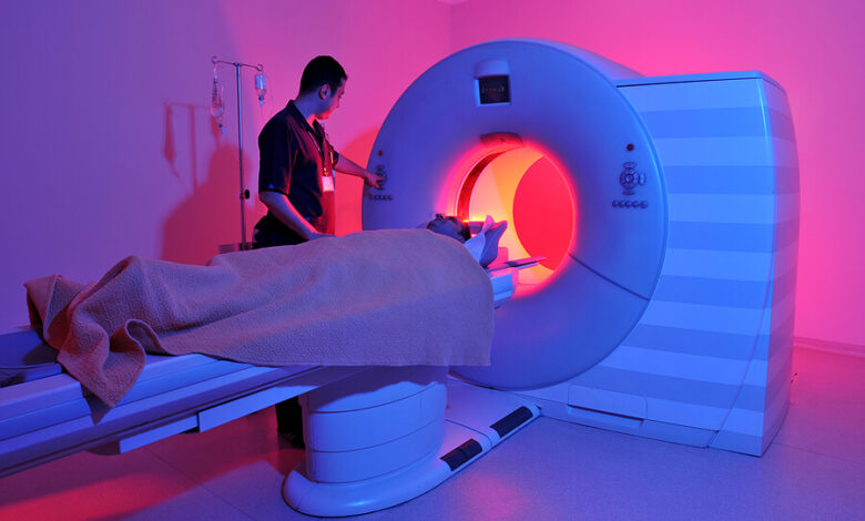
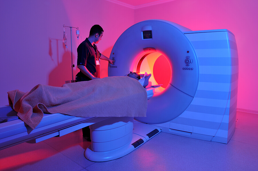
What is full body scan for detecting cancer and is it safe – What is a full body scan for detecting cancer and is it safe? That’s a question many people have, especially as advancements in medical imaging offer new ways to detect cancer earlier. This post dives into the world of full body scans – including PET, CT, and MRI – exploring how they work, their effectiveness, and importantly, the potential risks involved.
We’ll look at what each scan entails, compare their safety profiles, and discuss when a full body scan might be recommended by your doctor.
We’ll cover the different types of full body scans used to detect cancer, explaining their underlying principles and how they help doctors identify cancerous tissues. We’ll also delve into the interpretation of results, the limitations of these scans, and how they compare to more targeted screening methods. Ultimately, understanding the benefits and drawbacks of full body scans empowers you to make informed decisions about your health.
Introduction to Full Body Cancer Scans: What Is Full Body Scan For Detecting Cancer And Is It Safe
Full body scans play a crucial role in the early detection and diagnosis of cancer, significantly improving treatment outcomes and survival rates. These advanced imaging techniques allow doctors to visualize the entire body, identifying potential cancerous growths that might otherwise go undetected through more localized examinations. While not a replacement for other diagnostic methods, full body scans are a powerful tool in the fight against cancer.Different types of full body scans utilize varying principles to detect cancerous tissues.
They differ in their sensitivity and specificity, meaning how well they identify cancer and how well they avoid false positives. The choice of scan depends on several factors, including the type of cancer suspected, the patient’s medical history, and the doctor’s clinical judgment.
Types of Full Body Cancer Scans and Their Mechanisms
Several imaging modalities are employed for full body cancer scans, each operating on different physical principles. These scans provide complementary information, allowing for a more comprehensive assessment.PET (Positron Emission Tomography) scans use a radioactive tracer that’s injected into the bloodstream. Cancer cells, due to their high metabolic rate, absorb more of this tracer than healthy cells. The scan then detects the emitted radiation, creating a 3D image highlighting areas of increased metabolic activity, which are often indicative of cancerous tumors.
The resulting image shows areas of high tracer uptake as bright spots, helping to pinpoint the location and size of potential tumors. A PET scan is particularly useful in staging cancer, determining the extent of its spread.CT (Computed Tomography) scans use X-rays to create detailed cross-sectional images of the body. A CT scan can detect abnormalities in tissue density, such as tumors, which appear as areas of different density compared to surrounding healthy tissues.
So, you’re wondering about full body scans for cancer detection and their safety? It’s a complex issue with varying opinions on the risks and benefits. Interestingly, early detection is key in many diseases, and research even suggests that a simple eye test, as discussed in this fascinating article, can eye test detect dementia risk in older adults , might offer insights into dementia risk.
This highlights the importance of preventative health checks, prompting us to consider the role of full body scans in a broader preventative health strategy.
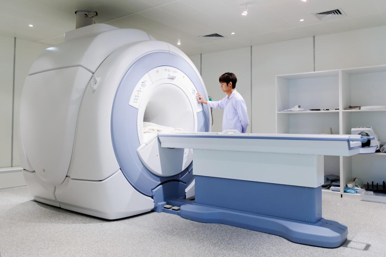
While not as specific as a PET scan in identifying cancerous tissue, CT scans offer excellent anatomical detail, making them useful for visualizing the size, shape, and location of tumors. CT scans are often used in conjunction with other imaging techniques, such as PET scans.MRI (Magnetic Resonance Imaging) scans use powerful magnets and radio waves to create detailed images of the body’s internal structures.
MRI is particularly sensitive to differences in water content within tissues. Cancerous tissues often have different water content compared to healthy tissues, allowing MRI to detect tumors that may not be visible on other scans. MRI is excellent for visualizing soft tissues and is often used to assess the extent of tumor involvement in organs and surrounding structures.
It’s particularly useful for brain and spinal cord cancers.
Specific Cancer Detection Methods within Full Body Scans
Full body scans aren’t a single test but a combination of imaging techniques, each offering unique insights into different aspects of the body’s internal structure. Understanding how these scans work and their strengths and weaknesses is crucial for accurate cancer detection. This section delves into the specific methods employed by common full-body scan components: PET, CT, MRI, and ultrasound.
So, you’re wondering about full body scans for cancer detection and their safety? It’s a complex issue, with different scans having different risks and benefits. Thinking about managing health concerns in children reminds me of the challenges faced by families dealing with conditions like Tourette Syndrome; finding effective strategies is key, and you can check out some helpful tips on managing this in children here: strategies to manage tourette syndrome in children.
Returning to full body scans, remember to always discuss any concerns with your doctor before undergoing any medical procedure.
PET Scan Technology
PET scans, or Positron Emission Tomography scans, utilize radioactive tracers to detect cancerous cells. These tracers, injected into the bloodstream, are absorbed at higher rates by rapidly dividing cells, such as those found in tumors. The tracer emits positrons, which collide with electrons, producing gamma rays that are detected by the PET scanner. The resulting images highlight areas of increased metabolic activity, indicating potential cancerous growths.
The higher the concentration of the tracer, the more likely the area is cancerous. This makes PET scans particularly useful in detecting the spread of cancer (metastasis) and assessing the effectiveness of treatment.
Comparison of Imaging Techniques
The following table compares PET, CT, MRI, and ultrasound scans across key parameters:
| Scan Type | Radiation Exposure | Image Resolution | Cost | Best Suited For |
|---|---|---|---|---|
| PET | Moderate (due to radioactive tracer) | Moderate | High | Detecting the spread of cancer, assessing treatment response; lymphomas, lung cancer, melanoma |
| CT | High (due to X-rays) | High | Moderate | Detecting bone and soft tissue abnormalities; lung cancer, colon cancer, pancreatic cancer |
| MRI | None | High | High | Imaging soft tissues; brain tumors, breast cancer, prostate cancer |
| Ultrasound | None | Moderate | Low | Imaging soft tissues and organs; breast cancer, thyroid cancer, ovarian cancer |
CT Scan Technology
CT scans, or Computed Tomography scans, utilize X-rays to generate detailed cross-sectional images of internal organs and structures. A rotating X-ray tube and detectors capture multiple images from different angles, which are then processed by a computer to create detailed 3D images. CT scans are particularly effective in visualizing bone and soft tissue structures, making them valuable for detecting tumors in various locations.
So, you’re wondering about full body scans for cancer detection and their safety? It’s a complex issue, with the technology constantly evolving. Understanding the risks is crucial, and sometimes, those risks mirror other health concerns. For example, high blood pressure is a major risk factor for stroke, and checking out this article on risk factors that make stroke more dangerous highlights how preventable some of these are.
Similarly, early detection of cancer through full body scans can save lives, but the radiation exposure needs careful consideration. The benefits and drawbacks must be weighed carefully with your doctor.
The high resolution allows for precise identification of tumor size, location, and potential spread to nearby tissues or lymph nodes.
MRI Scan Technology
MRI scans, or Magnetic Resonance Imaging scans, employ powerful magnetic fields and radio waves to create detailed images of the body’s internal structures. The magnetic field aligns the protons in the body’s water molecules, and radio waves are used to disrupt this alignment. As the protons return to their original alignment, they emit signals that are detected by the MRI scanner.
These signals are then processed to generate high-resolution images of soft tissues, making MRI particularly useful for detecting tumors in organs such as the brain, breast, and prostate. MRI excels at differentiating between different types of tissues, providing crucial information about tumor characteristics.
Examples of Cancer Detection Using Full Body Scans
Lung cancer detection often involves a combination of CT scans (to visualize the tumor and its size) and PET scans (to assess the extent of the disease and whether it has spread). Breast cancer detection may utilize mammography, ultrasound, and MRI, with MRI providing detailed images of breast tissue to identify the size and location of tumors. Colon cancer screening often involves CT colonography, a non-invasive CT scan technique that allows for visualization of the colon without the need for a colonoscopy.
The choice of specific imaging techniques depends on the type of cancer suspected, the patient’s medical history, and the stage of the disease.
Safety Aspects of Full Body Cancer Scans
Full body cancer scans, while offering valuable diagnostic capabilities, do carry potential risks and side effects. Understanding these risks is crucial for informed decision-making, allowing individuals to weigh the benefits against the potential downsides. The safety profile varies depending on the specific scanning technique employed.
Radiation Exposure in Different Scanning Techniques
Different full body scans utilize varying levels of ionizing radiation. CT scans, for example, involve significantly higher radiation doses compared to MRI scans, which use powerful magnetic fields and radio waves instead. PET scans also use ionizing radiation, but in conjunction with a radioactive tracer, leading to a different radiation exposure profile. The following chart provides a simplified comparison:
| Scan Type | Radiation Level (relative) | Notes |
|---|---|---|
| CT Scan | High | Higher radiation dose compared to other methods. |
| PET Scan | Moderate | Radiation from both the scanner and the injected tracer. |
| MRI Scan | None | Uses magnetic fields and radio waves; no ionizing radiation. |
It’s important to note that these are relative comparisons and the actual radiation dose received varies depending on factors such as the specific scanner used, scan duration, and the patient’s size. The radiation levels are carefully managed by medical professionals to keep them as low as reasonably achievable (ALARA principle).
Allergic Reactions to Contrast Agents
Some full body scans, particularly CT and MRI scans, may involve the use of contrast agents. These agents are substances that enhance the visibility of certain structures or tissues within the body. While generally safe, contrast agents can cause allergic reactions in some individuals, ranging from mild skin rashes to more severe anaphylactic shock. Pre-scan screening for allergies and appropriate pre-medications are standard practice to mitigate this risk.
Patients with a history of allergies should inform their doctor before undergoing the scan.
Safety Precautions During the Scanning Procedure, What is full body scan for detecting cancer and is it safe
Several safety precautions are implemented to minimize risks during full body cancer scans. These include:
- Shielding: Lead aprons and other shielding materials may be used to protect sensitive organs from unnecessary radiation exposure, particularly during CT scans.
- Optimized Scan Parameters: Radiologists carefully select scan parameters (e.g., radiation dose, scan time) to minimize radiation exposure while ensuring diagnostic image quality.
- Monitoring: During the procedure, medical personnel closely monitor the patient for any adverse reactions or complications.
- Emergency Protocols: Hospitals and imaging centers have established emergency protocols to handle any unforeseen events or allergic reactions during or after the scan.
Long-Term Health Implications of Repeated Full Body Scans
Repeated exposure to ionizing radiation from CT scans and PET scans increases the cumulative radiation dose, potentially raising the risk of long-term health effects such as cancer. While the risk is generally low for a single scan, frequent scans, especially over a short period, should be carefully considered and justified by the clinical need. The benefits of the diagnostic information obtained must outweigh the potential risks associated with repeated radiation exposure.
MRI scans, lacking ionizing radiation, do not pose the same long-term risks associated with repeated procedures.
Interpreting Full Body Scan Results

Source: website-files.com
Interpreting the results of a full body scan, often a type of whole-body imaging like a PET/CT scan, is a complex process requiring the expertise of trained radiologists. These specialists analyze the images for abnormalities, comparing them to established norms and considering the patient’s medical history. The goal is to identify any areas of concern that may warrant further investigation.Radiologists meticulously examine the images, looking for deviations from the expected appearance of organs and tissues.
This involves assessing factors like size, shape, density, and metabolic activity (in the case of PET scans). The interpretation process isn’t simply about spotting a single anomaly; it’s about integrating all the visual information to create a comprehensive picture of the patient’s health.
Image Analysis Techniques
Radiologists utilize sophisticated software and their extensive knowledge of anatomy and pathology to analyze the images. They may employ techniques like contrast enhancement, three-dimensional reconstruction, and image fusion (combining images from different modalities) to better visualize and interpret subtle findings. The interpretation is a detailed and iterative process, often involving zooming in on specific areas of interest and comparing them to previous scans (if available) to track changes over time.
Indicators of Cancer on Full Body Scans
Several findings on a full body scan can raise suspicion of cancer. For example, an unusually large or irregularly shaped mass in an organ, such as the liver or lung, could be indicative of a tumor. Abnormal uptake of radioactive tracers (in PET scans) in a specific area might suggest increased metabolic activity associated with cancerous cells. Changes in the size or shape of lymph nodes could indicate the spread of cancer.
These are just examples, and the specific findings that raise concern will vary depending on the type of scan and the patient’s individual circumstances. It is crucial to understand that these findings are not definitive diagnoses.
The Role of Biopsy and Further Testing
A full body scan is a screening tool, not a diagnostic test. While it can identify suspicious areas, it cannot definitively confirm the presence of cancer. Further investigations are always necessary to confirm a diagnosis. The most common confirmatory test is a biopsy, which involves removing a small tissue sample for microscopic examination. Other tests, such as blood tests and further imaging studies (e.g., MRI, ultrasound), may also be employed to provide a more complete picture and guide treatment decisions.
For instance, a suspicious lung nodule identified on a CT scan would likely be followed up with a biopsy to determine whether it is cancerous or benign. The results of these additional tests are crucial for determining the appropriate course of action, from close monitoring to surgical intervention.
Limitations of Full Body Cancer Scans

Source: cliffordclinic.com
Full body cancer scans, while offering a comprehensive approach to cancer detection, are not a silver bullet. Their effectiveness is significantly impacted by various factors, leading to limitations in their ability to detect all cancers and accurately diagnose their stage. Understanding these limitations is crucial for managing expectations and making informed decisions regarding cancer screening.
Types of Cancer Difficult to Detect
Certain cancers, due to their location, size, or biological characteristics, are inherently more challenging to detect with full body scans. For example, cancers located in areas with high background radiation or dense tissue, such as the pancreas or certain brain regions, may be obscured by the surrounding structures, making their identification difficult even with advanced imaging techniques. Similarly, early-stage cancers, which are often small and haven’t yet spread, may be too small to be detected by current scanning technologies.
Cancers that develop slowly and don’t cause significant changes in tissue structure might also evade detection. Examples include some slow-growing lymphomas and early-stage prostate cancers.
Limitations of Specific Scanning Techniques
The sensitivity and specificity of different scanning techniques vary considerably. For instance, while PET scans are excellent at detecting metabolically active tumors, they are less effective at identifying small, slow-growing cancers. CT scans, on the other hand, provide high-resolution anatomical images but may miss cancers that haven’t caused significant structural changes. MRI scans excel in visualizing soft tissues, but their longer scan times and higher costs limit their widespread use as a primary screening tool.
The accuracy of each technique depends on factors such as the type of cancer, its location, and the expertise of the radiologist interpreting the images. A false positive result (indicating cancer when there is none) can lead to unnecessary anxiety and invasive procedures, while a false negative (missing a cancer that is present) can delay diagnosis and treatment.
Situations Where Full Body Scans May Not Be Effective
- Early-stage cancers: Small tumors may be too small to be detected by current imaging technology.
- Cancers in difficult-to-image locations: Cancers located in areas with high background radiation or dense tissue (e.g., pancreas, brain) may be obscured.
- Slow-growing cancers: Cancers that develop slowly and don’t cause significant changes in tissue structure might go undetected.
- Certain cancer types: Some cancers, like certain types of leukemia and lymphoma, may not be effectively detected by imaging scans.
- Limitations of technology: Current scanning technology may not be sensitive enough to detect all types of cancer at all stages.
- Individual patient factors: Factors such as body size, underlying medical conditions, and the presence of metallic implants can affect image quality and interpretation.
Full Body Scans vs. Targeted Cancer Screening
Choosing the right cancer screening method is a crucial decision, balancing the benefits of early detection with the potential risks and limitations of each approach. Full body scans, while offering a comprehensive overview, differ significantly from targeted screenings like mammograms or colonoscopies, each with its own strengths and weaknesses. Understanding these differences is vital for making informed healthcare choices.Full body scans, often involving techniques like PET or CT scans, aim to detect cancerous growths anywhere in the body.
Targeted screenings, on the other hand, focus on specific organs or tissues known to be at higher risk for particular cancers based on age, family history, or other risk factors. This targeted approach allows for more focused investigation and, in many cases, is more cost-effective and less prone to false positives.
Comparison of Full Body Scans and Targeted Screenings
Full body scans provide a broad overview, potentially identifying cancers in unexpected locations. However, this advantage comes with a higher chance of detecting incidental findings – abnormalities that aren’t necessarily cancerous but require further investigation, potentially leading to unnecessary anxiety and further testing. Targeted screenings, by contrast, are more precise, focusing on areas with a higher likelihood of cancer development.
This precision minimizes unnecessary investigations and reduces the risk of false positives, though it also means cancers in other areas might be missed.
Advantages and Disadvantages
The following table summarizes the advantages and disadvantages of each approach:
| Feature | Full Body Scan | Targeted Screening |
|---|---|---|
| Advantages | Detects cancers in unexpected locations; potentially earlier detection of multiple cancers. | Higher accuracy for specific cancers; lower radiation exposure; less expensive; fewer false positives. |
| Disadvantages | High cost; high radiation exposure; high rate of false positives; may lead to unnecessary further investigations and anxiety. | Only screens for specific cancers; may miss cancers outside the targeted area. |
Cost-Effectiveness and Efficacy
The cost-effectiveness and efficacy of full body scans versus targeted screenings vary significantly depending on the specific cancer type and individual risk factors. A full body scan is generally more expensive and may not be more effective than targeted screenings for common cancers like breast or colorectal cancer in low-risk individuals.
| Cancer Type | Full Body Scan (Cost-Effectiveness/Efficacy) | Targeted Screening (Cost-Effectiveness/Efficacy) |
|---|---|---|
| Breast Cancer | Low cost-effectiveness; efficacy varies depending on the stage of cancer. | High cost-effectiveness; high efficacy in early detection, especially with mammograms. |
| Colorectal Cancer | Low cost-effectiveness; efficacy limited compared to colonoscopy. | High cost-effectiveness; high efficacy with colonoscopy. |
| Lung Cancer | Potentially higher efficacy in early detection for high-risk individuals; cost-effectiveness debatable. | Low cost-effectiveness for general population screening; higher efficacy for high-risk individuals with low-dose CT scans. |
Note: The above table provides a general overview and specific cost-effectiveness and efficacy data may vary based on specific technologies, healthcare systems, and individual patient factors.
Circumstances for Full Body Scan Recommendation
Full body scans are generally not recommended for routine cancer screening in the general population due to the high cost, radiation exposure, and high rate of false positives. However, there are specific circumstances where a full body scan might be considered:* Individuals with a strong family history of multiple cancers.
- Patients experiencing unexplained symptoms that suggest widespread cancer.
- Cases where targeted screening has yielded inconclusive results.
- Monitoring of cancer treatment response in certain situations.
It’s crucial to discuss the risks and benefits of a full body scan with your doctor to determine if it’s appropriate for your individual circumstances. The decision should be based on a careful evaluation of your personal risk factors, medical history, and potential benefits versus the risks associated with the procedure.
Conclusion
So, is a full body scan for cancer detection safe? The answer, as with most medical procedures, is nuanced. While these scans offer valuable diagnostic tools, they do carry potential risks, primarily related to radiation exposure. The decision to undergo a full body scan should be made in consultation with your doctor, weighing the potential benefits against the risks based on your individual circumstances and health history.
Remember, targeted screenings often offer a more effective and less invasive approach for many cancers. This post aimed to equip you with the information needed to have a productive conversation with your healthcare provider about your cancer screening options.
FAQ Corner
How accurate are full body scans in detecting cancer?
Accuracy varies greatly depending on the type of scan, the cancer type, and the stage of the cancer. No scan is 100% accurate, and false positives and negatives can occur.
Can I request a full body scan from my doctor?
While you can discuss your concerns with your doctor, full body scans aren’t typically recommended as routine screenings for healthy individuals. They’re usually reserved for individuals with specific symptoms, risk factors, or a family history of cancer.
What should I expect during a full body scan?
The procedure itself varies depending on the type of scan. You’ll likely need to lie still on a table for a period of time. Some scans involve the injection of contrast dye, while others don’t. Your doctor or technician will explain the procedure in detail before you begin.
Are there alternative screening methods?
Yes, many targeted screening methods exist, such as mammograms for breast cancer, colonoscopies for colon cancer, and Pap smears for cervical cancer. These are often preferred over full body scans for specific cancers due to their higher accuracy and lower radiation exposure.
